User:David Hume/sandbox: Difference between revisions
imported>David Hume No edit summary |
No edit summary |
||
| (28 intermediate revisions by 2 users not shown) | |||
| Line 1: | Line 1: | ||
{{AccountNotLive}} | |||
{{systemic}} | |||
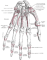
''' | The Triangular Bone (os triquetum; cuneiform bone) (Fig. 223).—The triangular bone may be distinguished by its pyramidal shape, and by an oval isolated facet for articulation with the pisiform bone. It is situated at the upper and ulnar side of the carpus. The superior surface presents a medial, rough, non-articular portion, and a lateral convex articular portion which articulates with the triangular articular disk of the wrist. The inferior surface, directed lateralward, is concave, sinuously curved, and smooth for articulation with the hamate. The dorsal surface is rough for the attachment of ligaments. The volar surface presents, on its medial part, an oval facet, for articulation with the pisiform; its lateral part is rough for ligamentous attachment. The lateral surface, the base of the pyramid, is marked by a flat, quadrilateral facet, for articulation with the lunate. The medial surface, the summit of the pyramid, is pointed and roughened, for the attachment of the ulnar collateral ligament of the wrist. 8 | ||
Articulations.—The triangular articulates with three bones: the lunate laterally, the pisiform in front, the hamate distally; and with the triangular articular disk which separates it from the lower end of the ulna. | |||
The Triangular Bone (os triquetrum; cuneiform bone) | |||
{{Image|triquetral.JPG|left|300px|triquetral}} | |||
{{Infobox Bone | | |||
Name = Triquetral bone | | |||
Latin = os triquetrum, os pyramidale, os triangulare | | |||
GraySubject = 54 | | |||
GrayPage = 224 | | |||
Image = Carpus.png | | |||
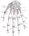
Caption = '''BONES OF HAND'''<BR>''Proximal:'' A=[[Scaphoid bone|Scaphoid]], B=[[Lunate bone|Lunate]], C=[[Triquetral bone|Triquetral]], D=[[Pisiform bone|Pisiform]]<BR>''Distal:'' E=[[Trapezium bone|Trapezium]], F=[[Trapezoid bone|Trapezoid]], G=[[Capitate bone|Capitate]], H=[[Hamate bone|Hamate]]<BR> | | |||
Image2 = Gray223.png | | |||
Caption2 = The left triquetal bone. | | |||
Origins = | | |||
Insertions = | | |||
Articulations = articulates with ''three'' bones:<BR>[[lunate]] laterally<BR>[[pisiform]] in front<BR>[[hamate]] distally<BR>triangular [[articular disk]] which separates it from the lower end of the [[ulna]]. | | |||
MeshName = Triquetrum+Bone | | |||
MeshNumber = A02.835.232.087.319.150.831 | | |||
DorlandsPre = o_07 | | |||
DorlandsSuf = 12598819 | | |||
}} | |||
The '''triquetral bone''' (also called '''triquetrum bone''', '''cuneiform bone''', '''pyramidal bone''', '''cubital bone''', '''three-cornered bone''', and '''triangular bone''') is located in the [[wrist]] on the medial side of the proximal row of the [[carpus]] between the [[lunate]] and [[pisiform]] bones. It is on the [[ulnar]] side of the hand, but does not articulate with the [[ulna]]. It connects with the [[pisiform]], [[hamate]], and [[lunate]] bones. It is the 3rd most commonly fractured carpal bone. | |||
The triangular bone may be distinguished by its pyramidal shape, and by an oval isolated facet for articulation with the pisiform bone. It is situated at the upper and ulnar side of the carpus. To facilitate its palpation in an exam, the hand must be radially deviated so that the triquetrium moves out from under the ulnar styloid process. The triquetrum may be difficult to find, since it also lies under the pisiform. | |||
The etymology derives from the Latin ''triquetrus'' which means "three-cornered." | |||
==Surfaces== | |||
The ''superior surface'' presents a medial, rough, non-articular portion, and a lateral convex articular portion which articulates with the triangular articular disk of the wrist. | |||
The ''inferior surface'', directed lateralward, is concave, sinuously curved, and smooth for articulation with the hamate. The dorsal surface is rough for the attachment of ligaments. | |||
The ''volar surface'' presents, on its medial part, an oval facet, for articulation with the pisiform; its lateral part is rough for ligamentous attachment. | |||
The ''lateral surface'', the base of the pyramid, is marked by a flat, quadrilateral facet, for articulation with the lunate. | |||
The ''medial surface'', the summit of the pyramid, is pointed and roughened, for the attachment of the ulnar collateral ligament of the wrist. | |||
== See also == | |||
*[[Bone#Terminology|Bone terminology]] | |||
*[[Terms for anatomical location]] | |||
==Additional images== | |||
<gallery> | |||
Image:Gray219.png|Bones of the left hand. Volar surface. | |||
Image:Gray220.png|Bones of the left hand. Dorsal surface. | |||
</gallery> | |||
{{Bones of upper extremity}} | |||
[[Category:Skeletal system]] | |||
[[Category:wrist]] | |||
{{musculoskeletal-stub}} | |||
[[fr:Os triquetrum]] | |||
[[nl:Os triquetrum]] | |||
[[sk:Trojhranná kosť]] | |||
By comparison, the term "palm" applies to only the palm (the grasping side) of the hand. The Romans actually used the word "palma" for the outstretched palm of the hand. | By comparison, the term "palm" applies to only the palm (the grasping side) of the hand. The Romans actually used the word "palma" for the outstretched palm of the hand. | ||
== | ==Topics in Anatomy== | ||
SYSTEMIC ANATOMY | |||
'''SYSTEMIC ANATOMY''' | |||
integumentary system | Introduction and Systemic Overview | ||
*Anatomical Nomenclature | |||
full skeleton | *Basic Structure and Function of Cells | ||
skull | *Integrating Cells into Tissues | ||
vertebral column | Systemic Overview | ||
ribcage | *Nervous System | ||
shoulder & arm | *Blood, Lymphoid Tissues and Haemopoiesis | ||
hand & wrist | *Functional Anatomy of the Musculoskeletal System | ||
pelvis | *Smooth Muscle and the Cardiovascular and Lymphatic systems | ||
leg & ankle | *Skin and its Appendages | ||
foot | *Endocrine System | ||
*Principles of Hormone Production and Secretion | |||
full body | *Embryology | ||
muscle histology | **Embryogenesis | ||
head & neck | **Prenatal and Neonatal Growth | ||
thorax | |||
shoulder & upper arm | [[Integumentary System]] | ||
*[[integumentary system]] | |||
forearm & hand | |||
abdomen | [[Skeletal System]] | ||
pelvis | *[[full skeleton]] | ||
leg & foot | *[[axial skeleton]] | ||
*[[skull]] | |||
nervous system | *[[vertebral column]] | ||
brain | *[[ribcage]] | ||
spinal cord | *[[appendicular skeleton]] | ||
autonomic nervous system | *[[shoulder & arm]] | ||
eye | *[[hand & wrist]] | ||
ear | *[[pelvis]] | ||
nose | *[[leg & ankle]] | ||
*[[foot]] | |||
endocrine | |||
hypothalamus & pituitary | [[Muscular System]] | ||
thyroid & parathyroids | *[[full body]] | ||
adrenal glands | *[[muscle histology]] | ||
pancreas | *[[head & neck]] | ||
ovaries | *[[thorax]] | ||
testes | *[[shoulder & upper arm]] | ||
*[[biceps brachii]] | |||
cardiovascular system | *[[forearm & hand]] | ||
lymphatic system | *[[abdomen]] | ||
*[[pelvis]] | |||
*[[thigh & knee]] | |||
*[[leg & foot]] | |||
[[Nervous System]] | |||
*[[nervous system]] | |||
*[[brain]] | |||
*[[spinal cord]] | |||
*[[autonomic nervous system]] | |||
*[[eye]] | |||
*[[ear]] | |||
*[[nose]] | |||
[[Endocrine System]] | |||
*[[endocrine system]] | |||
*[[hypothalamus & pituitary]] | |||
*[[thyroid & parathyroids]] | |||
*[[adrenal glands]] | |||
*[[pancreas]] | |||
*[[ovaries]] | |||
*[[testes]] | |||
[[Cardiovascular System]] ([[Circulatory System]]) | |||
*[[cardiovascular system]] | |||
*[[lymphatic system]] | |||
[[Lymphatic System]] | |||
[[Immune System]] | |||
[[Respiratory System]] | |||
*[[respiratory system]] | |||
*[[mouth]] | |||
*[[nose & throat]] | |||
*[[lung]] | |||
[[Digestive System]] | |||
*[[digestive system]] | |||
*[[alimentary canal]] | |||
*[[accessory organs]] | |||
*[[mouth & throat]] | |||
*[[esophagus & stomach]] | |||
*[[liver]] | |||
*[[gallbladder]] | |||
*[[pancreas & duodenum]] | |||
*[[small intestine]] | |||
*[[large intestine]] | |||
[[Reproductive System]] | |||
*[[male reproductive systerm]] | |||
*[[female reproductive system]] | |||
male | |||
female | |||
*[[Excretory System]] ([[Urinary System]]) | |||
*[[urinary system]] | |||
*[[kidneys]] | |||
Latest revision as of 03:43, 22 November 2023
The account of this former contributor was not re-activated after the server upgrade of March 2022.
The Triangular Bone (os triquetum; cuneiform bone) (Fig. 223).—The triangular bone may be distinguished by its pyramidal shape, and by an oval isolated facet for articulation with the pisiform bone. It is situated at the upper and ulnar side of the carpus. The superior surface presents a medial, rough, non-articular portion, and a lateral convex articular portion which articulates with the triangular articular disk of the wrist. The inferior surface, directed lateralward, is concave, sinuously curved, and smooth for articulation with the hamate. The dorsal surface is rough for the attachment of ligaments. The volar surface presents, on its medial part, an oval facet, for articulation with the pisiform; its lateral part is rough for ligamentous attachment. The lateral surface, the base of the pyramid, is marked by a flat, quadrilateral facet, for articulation with the lunate. The medial surface, the summit of the pyramid, is pointed and roughened, for the attachment of the ulnar collateral ligament of the wrist. 8
Articulations.—The triangular articulates with three bones: the lunate laterally, the pisiform in front, the hamate distally; and with the triangular articular disk which separates it from the lower end of the ulna.
The Triangular Bone (os triquetrum; cuneiform bone)

Template:Infobox Bone The triquetral bone (also called triquetrum bone, cuneiform bone, pyramidal bone, cubital bone, three-cornered bone, and triangular bone) is located in the wrist on the medial side of the proximal row of the carpus between the lunate and pisiform bones. It is on the ulnar side of the hand, but does not articulate with the ulna. It connects with the pisiform, hamate, and lunate bones. It is the 3rd most commonly fractured carpal bone.
The triangular bone may be distinguished by its pyramidal shape, and by an oval isolated facet for articulation with the pisiform bone. It is situated at the upper and ulnar side of the carpus. To facilitate its palpation in an exam, the hand must be radially deviated so that the triquetrium moves out from under the ulnar styloid process. The triquetrum may be difficult to find, since it also lies under the pisiform.
The etymology derives from the Latin triquetrus which means "three-cornered."
Surfaces
The superior surface presents a medial, rough, non-articular portion, and a lateral convex articular portion which articulates with the triangular articular disk of the wrist.
The inferior surface, directed lateralward, is concave, sinuously curved, and smooth for articulation with the hamate. The dorsal surface is rough for the attachment of ligaments.
The volar surface presents, on its medial part, an oval facet, for articulation with the pisiform; its lateral part is rough for ligamentous attachment.
The lateral surface, the base of the pyramid, is marked by a flat, quadrilateral facet, for articulation with the lunate.
The medial surface, the summit of the pyramid, is pointed and roughened, for the attachment of the ulnar collateral ligament of the wrist.
See also
Additional images
Bones of the left hand. Volar surface.
Bones of the left hand. Dorsal surface.


Template:Bones of upper extremity
fr:Os triquetrum nl:Os triquetrum sk:Trojhranná kosť
By comparison, the term "palm" applies to only the palm (the grasping side) of the hand. The Romans actually used the word "palma" for the outstretched palm of the hand.
Topics in Anatomy
SYSTEMIC ANATOMY Introduction and Systemic Overview
- Anatomical Nomenclature
- Basic Structure and Function of Cells
- Integrating Cells into Tissues
Systemic Overview
- Nervous System
- Blood, Lymphoid Tissues and Haemopoiesis
- Functional Anatomy of the Musculoskeletal System
- Smooth Muscle and the Cardiovascular and Lymphatic systems
- Skin and its Appendages
- Endocrine System
- Principles of Hormone Production and Secretion
- Embryology
- Embryogenesis
- Prenatal and Neonatal Growth
- full skeleton
- axial skeleton
- skull
- vertebral column
- ribcage
- appendicular skeleton
- shoulder & arm
- hand & wrist
- pelvis
- leg & ankle
- foot
- full body
- muscle histology
- head & neck
- thorax
- shoulder & upper arm
- biceps brachii
- forearm & hand
- abdomen
- pelvis
- thigh & knee
- leg & foot
- endocrine system
- hypothalamus & pituitary
- thyroid & parathyroids
- adrenal glands
- pancreas
- ovaries
- testes