File:Plaque assay macro.jpg: Difference between revisions
Jump to navigation
Jump to search
imported>Nancy Sculerati (Macroscopic photograph of viral plaque formation. Vero cells, which grew confluently on the bottom of the plastic plate (1.5 cm diameter), were infected with herpes simplex virus (approximately 75 plaque forming unit), and then cultured over night to make viral plaque. Living cells were stained with crystal violet. The viral plaques, each was from one virion, remained transparent. uploaded from Wikiedia commons I, the author of this work, hereby publish it under the following licenses: Permi...) |
imported>Joe Quick (license template) |
||
| Line 1: | Line 1: | ||
Macroscopic photograph of viral plaque formation. Vero cells, which grew confluently on the bottom of the plastic plate (1.5 cm diameter), were infected with herpes simplex virus (approximately 75 plaque forming unit), and then cultured over night to make viral plaque. Living cells were stained with crystal violet. The viral plaques, each was from one virion, remained transparent. | Macroscopic photograph of viral plaque formation. Vero cells, which grew confluently on the bottom of the plastic plate (1.5 cm diameter), were infected with herpes simplex virus (approximately 75 plaque forming unit), and then cultured over night to make viral plaque. Living cells were stained with crystal violet. The viral plaques, each was from one virion, remained transparent. | ||
uploaded from Wikiedia commons | uploaded from Wikiedia commons | ||
I, the author of this work, hereby publish it under the following licenses: | I, the author of this work, hereby publish it under the following licenses:{{GFDL}} | ||
Revision as of 11:43, 4 May 2007
Macroscopic photograph of viral plaque formation. Vero cells, which grew confluently on the bottom of the plastic plate (1.5 cm diameter), were infected with herpes simplex virus (approximately 75 plaque forming unit), and then cultured over night to make viral plaque. Living cells were stained with crystal violet. The viral plaques, each was from one virion, remained transparent. uploaded from Wikiedia commons I, the author of this work, hereby publish it under the following licenses:Template:GFDL
File history
Click on a date/time to view the file as it appeared at that time.
| Date/Time | Thumbnail | Dimensions | User | Comment | |
|---|---|---|---|---|---|
| current | 19:56, 11 March 2022 | 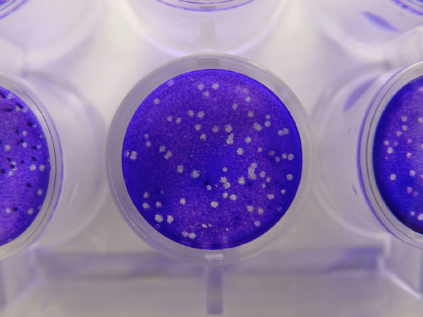 | 600 × 450 (43 KB) | Maintenance script (talk | contribs) | == Summary == Importing file |
You cannot overwrite this file.
File usage
The following 2 files are duplicates of this file (more details):
- File:Plaque assay macro.jpg from a shared repository
- File:Plaque assay macro.jpg from Wikimedia Commons
The following page uses this file: