File:Brain morphometry image segmentation.png

Original file (2,937 × 1,776 pixels, file size: 1.18 MB, MIME type: image/png)
Summary
| Title / Description
|
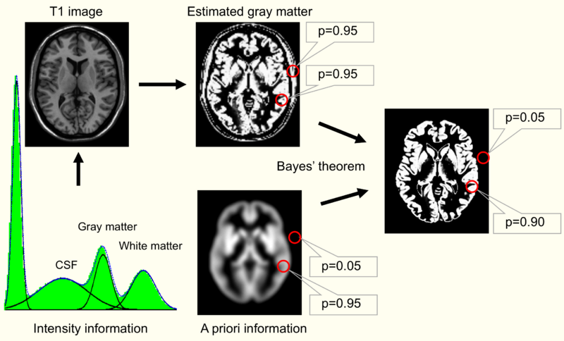
Image segmentation using a priori information. In the first step, the image intensities of the T1 image (upper left) are used to plot their frequencies in a histogram. Several peaks – corresponding to different image intensities of the tissue classes – can be differentiated. In the next step, Gaussian curves for each tissue class are fitted into the histogram to estimate the probability of a voxel belonging to that tissue class (bottom left). A map for gray matter is shown (upper right) with the estimated probability for two selected locations (red circles). Based solely on a similar image intensity, the cerebral and the extracranial spot exhibit a similar probability for belonging to gray matter. This can be corrected by combining the image intensity-based information with prior information (below), e.g. using a Bayesian approach. |
|---|---|
| Author(s)
|
Daniel Mietchen and Christian Gaser |
| Copyright holder
|
Please edit this page and add the name of the copyright holder, or note why this is not applicable. |
| Source
|
Please edit this page and add the source of this media, or note that it is unknown. |
| Date created
|
2009 |
| Country of first publication
|
Please edit this page and add the country of first publication, or note that it is unknown. |
| Notes
|
You can edit this page and add notes here which may be useful to people who wish to re-use this media. |
| Other versions
|
If there are other versions of this media on CZ, please list them here. |
| Using this image on CZ
|
Copy the code below to add this image to a Citizendium article, changing the size, alignment, and caption as necessary.
|
Please send email to manager A T citizendium.org .
Licensing/Copyright status
This media, Brain morphometry image segmentation.png, is licenced under the Creative Commons Attribution-ShareAlike 3.0 Unported License
You are free:
To Share — To copy, distribute and transmit the work; To Remix — To adapt the work.
Under the following conditions:
Attribution — You must attribute the work in the manner specified by the author or licensor (but not in any way that suggests that they endorse you or your use of the work). Share Alike — If you alter, transform, or build upon this work, you may distribute the resulting work only under the same, similar or a compatible licence.
For any reuse or distribution, you must make clear to others the licence terms of this work (the best way to do this is with a link to this licence's web page). Any of the above conditions can be waived if you get permission from the copyright holder. Nothing in this licence impairs or restricts the author's moral rights.
Read the full licence.
File history
Click on a date/time to view the file as it appeared at that time.
| Date/Time | Thumbnail | Dimensions | User | Comment | |
|---|---|---|---|---|---|
| current | 19:53, 11 March 2022 | | 2,937 × 1,776 (1.18 MB) | Maintenance script (talk | contribs) | == Summary == Importing file |
You cannot overwrite this file.
File usage
The following 2 pages use this file: